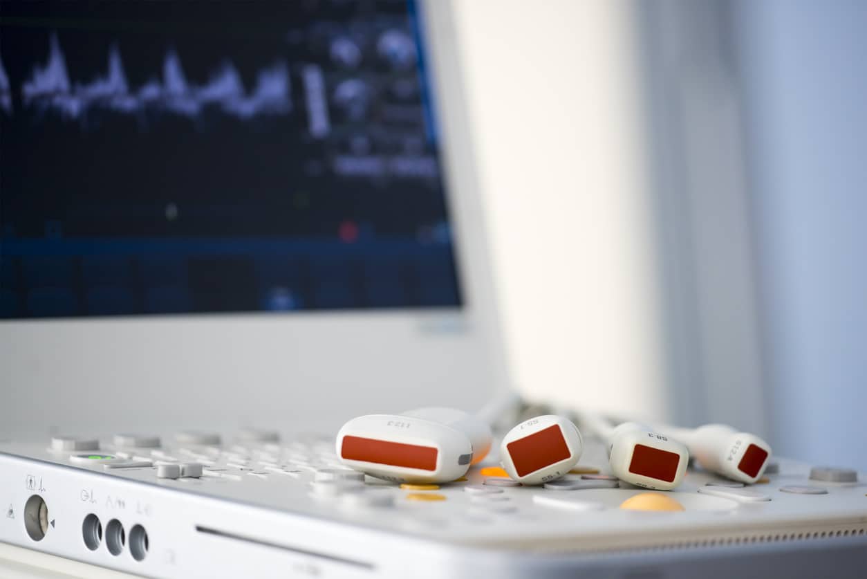
What is Vein Ultrasound?
Vein Ultrasound imaging, or sonography, is a procedure that produces images of the inside of the body using high-frequency sound waves. These images are captured in real-time and are able to show the structure and movement of blood flow.
What is Vein Ultrasound used for at the Los Angeles Vein Center?
Vein Ultrasound imaging can be used to monitor and diagnose a wide range of conditions. In particular, it is effective at evaluating the location and severity of venous insufficiency. It can also detect deep vein thrombosis and is an important tool during varicose vein procedures. At the Los Angeles Vein Center, we offer in-office Ultrasound imaging to help with diagnosis and procedure.

This leads to fast and effective treatment of various venous disorders. Dr. Lee has been credentialed by the American Registry for Diagnostic Medical Sonography (ARDMS) and is a Registered Physician in Vascular Interpretation (RPVI). In addition, our ultrasounds are performed by a Registered Vascular Technologist (RVT) credentialed by ARDMS and a Registered Vascular Specialist credentialed by Cardiovascular Credentialing International (CCI).
Candidates for Vein Ultrasound
If you’re showing signs or symptoms of decreased blood flow in the arteries or veins of your legs, arms, or neck, your doctor could order a vein ultrasound. A reduced amount of blood flow may be due to a blockage in the artery, a blood clot inside a blood vessel, or an injury to a blood vessel. Vein ultrasound can estimate the blood flow through your blood vessels.
These are conditions that could merit your doctor requesting a vein ultrasound:
- Deep vein thrombosis — This is a condition that occurs when a blood clot forms in a vein deep inside your body (usually in the legs or hips)
- Superficial thrombophlebitis — This is an inflammation of the veins due to a blood clot in a vein just below the skin’s surface.
- Arteriosclerosis — This is a narrowing or hardening of the arteries that supply blood to the legs and feet.
- Thromboangiitis obliterans — This is a rare disease where the blood vessels of the hands and feet become inflamed and swollen.
- Vascular tumors in your arms or legs
Benefits of a Vein Ultrasound
A vein ultrasound is a non-invasive, simple procedure that can be used to diagnose many different conditions by producing images of the soft tissues, which often do not show up well on x-rays. There is no ionizing radiation used during this procedure and is a proven safe diagnostic tool.
How accurate is Vein Ultrasound?
According to the National Blood Clot Alliance, a vein ultrasound finds about 95 percent of deep vein thrombosis clots in the large veins above the knee. Venous ultrasound identifies only about 60 to 70 percent of deep vein thrombosis in calf veins. These clots are less likely to become pulmonary embolisms than those that form above the knee.
How should I prepare for a Vein Ultrasound procedure at L.A. Vein Center?
There aren’t any preparations needed unless we’re looking at veins in your abdomen (which is quite rare). In that case, you would need to fast for a period of time.
Most of these procedures at L.A. Vein don’t require any preparation.
Can I exercise before getting a Vein Ultrasound?
Yes. But you should provide at least an hour for your body to return to normal stress levels before your vein ultrasound.
The Vein Ultrasound Procedure
During the ultrasound, the patient is positioned on an examination table and a clear gel is applied to the area being examined. A transducer is then firmly pressed against the skin and swept back and forth to record the sound waves that are used to obtain the image. The image appears on a computer screen in real-time, so that the patient and doctor can view it together.

How long does a Vein Ultrasound take?
Ultrasound tests usually take between 30 and 45 minutes.
What our patients have to say
"I have been seeing this team of incredibly warm and friendly professionals all year to work on my veins, and can’t even begin to recommend them highly enough. Dr. Lee is kind, compassionate, and extremely competent in her field. It seems like she really knows how to hire too because the entire staff is capable and proficient in their jobs. There is never a long wait and with every visit I see progress and feel less pain. My results have been very noticeable and very worth it!"
Is there any discomfort during a Vein Ultrasound?
There is little if any discomfort during a vein ultrasound. The transducer is pressed against your skin and moved along your arm or leg. The procedure is simple.
Can Vein Ultrasound tests show blood clots?
Yes. Finding clots is one of the main purposes of these imaging tests. Clots occur in a deep vein in the legs or the hip area. Duplex ultrasound processes are used. In the first part, brightness modulation ultrasound (also known as B-mode ultrasound) is used to obtain an image or picture. Once the area with the potential clot is defined, the sonographer (the operator of the ultrasound machine) tries to collapse or compress the veins. If a vein cannot be compressed because it has a clot, then the diagnosis of deep vein thrombosis is made.
The second phase of duplex ultrasound is used to detect abnormalities of blood flow. Sound waves are bounced off the blood within a vein. Flowing blood changes the sound waves by the Doppler effect. The ultrasound machine can detect these changes and determine whether the blood inside a vein is flowing normally. If there is an absence of blood flow, the diagnosis of deep vein thrombosis is confirmed.
Who interprets the results from my Vein Ultrasound?
Dr. Lee interprets your vein ultrasound. Board-certified in both vascular surgery and general surgery, Dr. Lee has unique, extensive experience with the vascular system and treatment of venous disease. Dr. Lee has also been credentialed by the American Registry for Diagnostic Medical Sonography and is a Registered Physician in Vascular Interpretation.
How will I discuss my results with my doctor?
Dr. Lee may discuss your results with you after your venous ultrasound, or she may send the results to your doctor who requested the test. Your doctor will then share your results with you.
Your vein ultrasound may create the need for a follow-up test or exam, as a potential abnormality could merit more evaluation or a special imaging technique, such as an MRI.
Risks of a Vein Ultrasound
Ultrasound waves have no harmful effects on humans. There are no risks inherent in these tests. At times the clot may not be able to be identified, especially if the clot is below the knee in the calf. But this really isn’t a risk pertaining to the test itself.
Schedule a Consultation
Contact our Los Angeles office today at (818) 325-0400 to learn more about our Vein Ultrasound services, or to make an appointment.

